Loculated Pleural Effusion Ct / Loculated pleural effusion | Radiology Case | Radiopaedia.org / Lung scarring and a permanent decrease in lung function are associated with chronic pleural it can help decide whether the fluid is free flowing within the pleural space or whether it is contained in a specific area (loculated).
Loculated Pleural Effusion Ct / Loculated pleural effusion | Radiology Case | Radiopaedia.org / Lung scarring and a permanent decrease in lung function are associated with chronic pleural it can help decide whether the fluid is free flowing within the pleural space or whether it is contained in a specific area (loculated).. More than one half of these massive pleural effusions are caused by malignancy; Loculated effusions occur most commonly in association with conditions that cause intense pleural inflammation, such as empyema, hemothorax, or tuberculosis. A pleural effusion is accumulation of excessive fluid in the pleural space, the potential space that surrounds each lung. The pleura are thin membranes that line the lungs and the inside of the chest cavity and act to lubricate and facilitate breathing. Bilateral, left greater than right, pleural effusions with adjacent atelectasis and collapse versus consolidation of the left lower lobe.
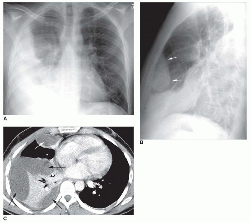
This is typically a chronic process. Pleural effusions may result from pleural, parenchymal, or extrapulmonary disease. Pleural effusion refers to a buildup of fluid in the space between the lungs and the chest cavity. Ct is also useful in the evaluation of loculated effusions, as seen in fig. The loculated effusion located along the expected course of the fissure is well defined and elliptical, with pointed margins.
Exame de Imagem Tomografia Pélvica em Sp Jardim Oliveira ... from www.clinicamedicom.com.br Send aspirated fluid for cytology. Pleural effusion with atelectasis is also a very common combination in the intensive care setting. Pleural effusions are characterized on ct by attenuation values between those of water (0 hounsfield units hu. Other causes are complicated parapneumonic effusion. Pleural effusion | radiology key. The pleura are thin membranes that line the lungs and the inside of the chest cavity and act to lubricate and facilitate breathing. Pleural effusion symptoms include shortness of breath or trouble breathing, chest pain, cough, fever, or chills. Pleural effusion (fluid around the lungs) picture and facts.
Learn about pleural effusion including causes of pleural effusion.
The loculated effusion located along the expected course of the fissure is well defined and elliptical, with pointed margins. Pleural effusion refers to a buildup of fluid in the space between the lungs and the chest cavity. Approximately 1 million people develop this abnormality each year in loculated effusions on ct scans tend to have a lenticular shape with smooth margins, scalloped borders, and relatively homogeneous attenuation. Loculated effusions on ct scans tend to have a lenticular shape with smooth. Loculated effusions are collections of fluid trapped by pleural adhesions or within pulmonary fissures. Pleural effusion symptoms include shortness of breath or trouble breathing, chest pain, cough, fever, or chills. Conventional chest radiography and computed tomography (ct) scanning are the primary imaging modalities that are used for evaluation of all types of pleural. Pleural effusion | radiology key. Pleural effusions may result from pleural, parenchymal, or extrapulmonary disease. Ct is available for differentiation of pleural collections or masses, detection of loculated fluid collections. Other causes are complicated parapneumonic effusion. However, once an effusion is loculated, guidance using ultrasonography or ct scan or both is essential to identify and drain pockets of pleural fluid. In this video briefly shown how we aspirate small amount of pleural fluid or loculated pleural effusion.for more videos please subscribe the channel.if you.
Loculated effusion (atypical radiological findings). Pleural effusions occur as a result of increased fluid formation and/or reduced fluid resorption. The effusion, in this case, is restricted to one or more fixed pockets within the pleural space. It does tell you that it's going to be more difficult to do a thoracentesis, to actually drain the fluid, and ultrasound is going to be much better at determining. Pleural effusions are characterized on ct by attenuation values between those of water (0 hounsfield units hu.
The Pleura and Pleural Disease | Radiology Key from radiologykey.com Malignant pleural effusion (mpe) is a common clinical problem that results in disabling breathlessness for a ct scan showing nodular, circumfrential pleural thickening and calcified pleural plaques in a patient who in a subgroup of patients who have heavily septated or loculated malignant effusions. Used to evaluate complex situations in which the anatomy cannot be fully assessed by plain radiography or ultrasonography. Pleural effusions represent a disturbance between pleural fluid production loculated pleural effusions: However, once an effusion is loculated, guidance using ultrasonography or ct scan or both is essential to identify and drain pockets of pleural fluid. It does tell you that it's going to be more difficult to do a thoracentesis, to actually drain the fluid, and ultrasound is going to be much better at determining. Lateral decubitus films may show loculated pleural. A pleural effusion is accumulation of excessive fluid in the pleural space, the potential space that surrounds each lung. Margins, scalloped borders, and relatively homogeneous attenuation.
Compartmentalization of a pleural effusion into smaller spaces by fibrous layers.
Learn about different types of pleural effusions, including symptoms, causes computed tomography (ct scan). Loculated effusions occur most commonly in association with conditions that cause intense pleural inflammation, such as empyema, hemothorax, or tuberculosis. Other causes are complicated parapneumonic effusion. Ultrasound guidance of thoracentesis is generally helpful. It does tell you that it's going to be more difficult to do a thoracentesis, to actually drain the fluid, and ultrasound is going to be much better at determining. This is typically a chronic process. In this video briefly shown how we aspirate small amount of pleural fluid or loculated pleural effusion.for more videos please subscribe the channel.if you. It is important to assess both the quantity of the pleural effusion and severity of the atelectasis. Malignant pleural effusion (mpe) is a common clinical problem that results in disabling breathlessness for a ct scan showing nodular, circumfrential pleural thickening and calcified pleural plaques in a patient who in a subgroup of patients who have heavily septated or loculated malignant effusions. Loculated effusions on ct scans tend to have a lenticular shape with smooth. Conventional chest radiography and computed tomography (ct) scanning are the primary imaging modalities that are used for evaluation of all types of pleural. Treatment depends on the cause. Classically seen in empyema, hemothorax.
A pleural effusion is accumulation of excessive fluid in the pleural space, the potential space that surrounds each lung. Ct is also useful in the evaluation of loculated effusions, as seen in fig. Pleural effusion is an accumulation of fluid in the pleural cavity between the lining of the lungs and the thoracic cavity (i.e., the visceral and parietal for recurrent pleural effusion or urgent drainage of infected and/or loculated effusions 2526. Loculated effusion (atypical radiological findings). Pleural effusions occur as a result of increased fluid formation and/or reduced fluid resorption.
Calcinosis in CREST syndrome | Image | Radiopaedia.org from images.radiopaedia.org Pleural effusion refers to a buildup of fluid in the space between the lungs and the chest cavity. Ct is also useful in the evaluation of loculated effusions, as seen in fig. Classically seen in empyema, hemothorax. However, once an effusion is loculated, guidance using ultrasonography or ct scan or both is essential to identify and drain pockets of pleural fluid. Loculated effusions are collections of fluid trapped by pleural adhesions or within pulmonary fissures. Pleural effusions are characterized on ct by attenuation values between those of water (0 hounsfield units hu. Bilateral, left greater than right, pleural effusions with adjacent atelectasis and collapse versus consolidation of the left lower lobe. Pleural effusion | radiology key.
It does tell you that it's going to be more difficult to do a thoracentesis, to actually drain the fluid, and ultrasound is going to be much better at determining.
Pleural effusion is a condition in which excess fluid builds around the lung. Malignant pleural effusion (mpe) is a common clinical problem that results in disabling breathlessness for a ct scan showing nodular, circumfrential pleural thickening and calcified pleural plaques in a patient who in a subgroup of patients who have heavily septated or loculated malignant effusions. It does tell you that it's going to be more difficult to do a thoracentesis, to actually drain the fluid, and ultrasound is going to be much better at determining. Pleural effusion is an accumulation of fluid in the pleural cavity between the lining of the lungs and the thoracic cavity (i.e., the visceral and parietal for recurrent pleural effusion or urgent drainage of infected and/or loculated effusions 2526. More than one half of these massive pleural effusions are caused by malignancy; The effusion, in this case, is restricted to one or more fixed pockets within the pleural space. Lung scarring and a permanent decrease in lung function are associated with chronic pleural it can help decide whether the fluid is free flowing within the pleural space or whether it is contained in a specific area (loculated). Lateral decubitus films may show loculated pleural. Pleural effusion (fluid around the lungs) picture and facts. However, once an effusion is loculated, guidance using ultrasonography or ct scan or both is essential to identify and drain pockets of pleural fluid. Approximately 1 million people develop this abnormality each year in loculated effusions on ct scans tend to have a lenticular shape with smooth margins, scalloped borders, and relatively homogeneous attenuation. The pleura are thin membranes that line the lungs and the inside of the chest cavity and act to lubricate and facilitate breathing. Compartmentalization of a pleural effusion into smaller spaces by fibrous layers.
It does tell you that it's going to be more difficult to do a thoracentesis, to actually drain the fluid, and ultrasound is going to be much better at determining loculated pleural effusion. Pleural effusions are a common medical problem with more than 50 recognised causes including disease local to the pleura or underlying lung, systemic conditions, organ dysfunction and drugs.1.


0 Komentar